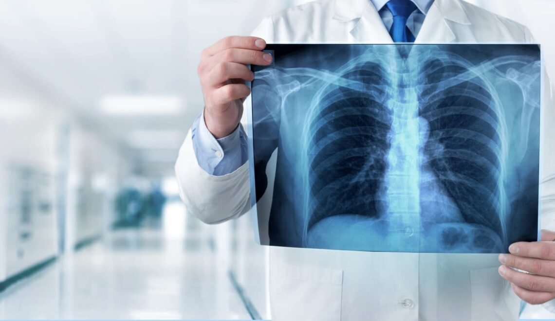
X-Ray
Intro:
The accidental discovery of X-ray took place in 1895 when Wilhelm Roentgen, a physics professor in Wurzburg, Bavaria, stumbled upon it during an experiment. Consequently, in 1896, the first radiology department was established at the Glasgow Royal Infirmary. Subsequently, X-ray machines found diverse applications, including the diagnosis of bone fractures and the localization of bullets during World War I. This single revelation brought a significant breakthrough to the field of medicine, revolutionizing it by enabling non-invasive visualization of internal structures within the human body without the need for surgical incisions and associated blood loss.
How does it work?
X-rays, like visible light, belong to the category of electromagnetic radiation. Nevertheless, X-rays possess higher energy levels and have the ability to penetrate various objects, including the human body. In the field of medicine, X-rays are employed to create visual representations of internal tissues and structures. When X-rays pass through the body and manage to reach an X-ray detector positioned on the opposite side of the patient, an image is produced. This image corresponds to the “shadows” formed by the objects present within the body.
Photographic film is one type of X-ray detector, although there exist numerous other detector varieties that are used for generating digital images. The resulting images from this process are commonly referred to as radiographs.
To initiate the creation of a radiograph, the patient is positioned in such a way that the specific part of their body intended for imaging is situated between an x-ray source and an x-ray detector. Once the machine is activated, x-rays traverse through the body encountering varying degrees of absorption by different tissues, depending on the radiological density of those tissues. This radiological density is governed by the density and atomic number of the material being imaged, denoting the number of protons in an atom’s nucleus.
For instance, our bones consist of calcium, which possesses a higher atomic number compared to most other tissues. This particular characteristic enables bones to easily absorb x-rays, consequently yielding a high level of contrast on the x-ray detector. Consequently, bony structures appear whiter than other tissues against the black background of a radiograph. On the other hand, tissues with lower radiological density, such as fat, muscle, and air-filled cavities like the lungs, allow x-rays to pass more effortlessly. As a result, an X-ray image represents these structures in different shades of gray.
When is it used?
X-ray radiography is capable of identifying a range of medical conditions and abnormalities, including bone fractures, specific tumors and abnormal masses, pneumonia, certain injuries, calcifications, foreign objects, and dental issues.
Different Procedures:
Mammography: A type of imaging technique involving radiography of the chest region, primarily utilized for the detection and diagnosis of cancer. On the radiograph, tumors typically appear as masses of regular or irregular shape, displaying a relatively brighter shade compared to the surrounding background (i.e., whiter on a black background or blacker on a white background). Additionally, mammograms have the ability to identify minuscule calcium deposits known as microcalcifications, visible as extremely bright specks in the mammogram. Although microcalcifications are generally harmless, specific formations of these tiny calcium bits could potentially indicate the presence of cancer.
Computed tomography, also known as CT, is a medical imaging technique that integrates conventional x-ray technology with computer processing capabilities. This integration enables the creation of a sequence of cross-sectional body images which can subsequently be merged to generate a three-dimensional x-ray representation. CT images offer higher levels of detail compared to regular radiographs, granting physicians the valuable capability to examine internal structures from numerous perspectives.
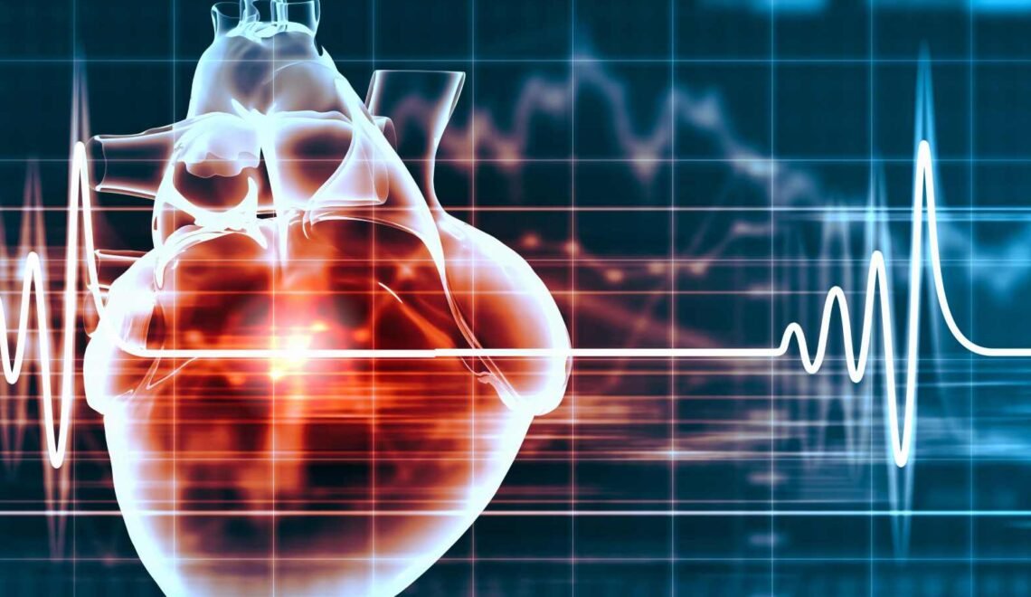
Fluoroscopy employs x-rays and a fluorescent screen to capture real-time images of bodily movements and diagnostic processes. It allows the tracking of contrast agents administered via injection or ingestion to follow their pathways accurately. For instance, fluoroscopy enables visualization of the heart’s beating motion and facilitates the observation of blood circulation in the heart muscles, blood vessels, and any other part of the body by utilizing radiographic contrast agents. Moreover, this technology assists in guiding a catheter with internal threading during cardiac angioplasty, a minimally invasive procedure used for clearing blockages in the heart’s blood supply arteries, with the aid of a radiographic contrast agent.
Therapeutic radiation therapy plays a crucial role in treating cancer by employing high-energy radiation, such as X-rays, to eradicate cancerous tumors and cells through DNA damage. It is essential to note that the radiation dosage administered for cancer treatment far exceeds the dosage used for diagnostic imaging. This therapeutic radiation can be delivered either externally through a machine positioned outside of the body or internally by employing radioactive substances placed inside or in close proximity to tumor cells, or even injected into the bloodstream.
Risks
When employed correctly, the advantages of utilizing x-ray scans for diagnostic purposes far outweigh the associated risks. X-ray scans have the capability to identify potentially perilous ailments like obstructed blood vessels, bone cancer, and infections. Nevertheless, it is important to acknowledge that x-rays emit ionizing radiation, a type of radiation that holds the potential to cause harm to living tissue. This risk escalates in tandem with the cumulative number of exposures an individual experiences throughout their lifetime. However, it is worth noting that the likelihood of developing cancer due to radiation exposure is generally minimal.
When an x-ray is performed on a pregnant woman, it does not pose any recognized risks to the baby as long as the specific area being examined does not involve the abdomen or pelvis. Typically, when imaging the abdomen and pelvis is required, healthcare professionals opt for radiation-free examinations like magnetic resonance imaging (MRI) or ultrasound. Nevertheless, if these methods fail to provide the necessary information, or if there is a time constraint or emergency situation, an x-ray can be considered as a suitable alternative for imaging purposes.
Children have an increased susceptibility to ionizing radiation and a more extended lifespan, which results in a comparatively greater chance of developing cancer from exposure to this type of radiation in comparison to adults. Concerned parents may consider inquiring with the technologist or doctor whether the machine settings have been appropriately tailored for the use on children.